Beranda
/ Computed Axial Tomography Cat - Computed Axial Tomography (CAT) Scan on Make a GIF / Computerized axial tomography (cat) adalah sebuah penggambaran medis yang menggunakan tomografi melalui proses geometri yang menghasilkan gambar tiga dimensi dari bagian dalam sebuah objek dari satu seri besar gambar sinar x dua dimensi dalam satu putaran axis
Computed Axial Tomography Cat - Computed Axial Tomography (CAT) Scan on Make a GIF / Computerized axial tomography (cat) adalah sebuah penggambaran medis yang menggunakan tomografi melalui proses geometri yang menghasilkan gambar tiga dimensi dari bagian dalam sebuah objek dari satu seri besar gambar sinar x dua dimensi dalam satu putaran axis
Insurance Gas/Electricity Loans Mortgage Attorney Lawyer Donate Conference Call Degree Credit Treatment Software Classes Recovery Trading Rehab Hosting Transfer Cord Blood Claim compensation mesothelioma mesothelioma attorney Houston car accident lawyer moreno valley can you sue a doctor for wrong diagnosis doctorate in security top online doctoral programs in business educational leadership doctoral programs online car accident doctor atlanta car accident doctor atlanta accident attorney rancho Cucamonga truck accident attorney san Antonio ONLINE BUSINESS DEGREE PROGRAMS ACCREDITED online accredited psychology degree masters degree in human resources online public administration masters degree online bitcoin merchant account bitcoin merchant services compare car insurance auto insurance troy mi seo explanation digital marketing degree floridaseo company fitness showrooms stamfordct how to work more efficiently seowordpress tips meaning of seo what is an seo what does an seo do what seo stands for best seotips google seo advice seo steps, The secure cloud-based platform for smart service delivery. Safelink is used by legal, professional and financial services to protect sensitive information, accelerate business processes and increase productivity. Use Safelink to collaborate securely with clients, colleagues and external parties. Safelink has a menu of workspace types with advanced features for dispute resolution, running deals and customised client portal creation. All data is encrypted (at rest and in transit and you retain your own encryption keys. Our titan security framework ensures your data is secure and you even have the option to choose your own data location from Channel Islands, London (UK), Dublin (EU), Australia.
Computed Axial Tomography Cat - Computed Axial Tomography (CAT) Scan on Make a GIF / Computerized axial tomography (cat) adalah sebuah penggambaran medis yang menggunakan tomografi melalui proses geometri yang menghasilkan gambar tiga dimensi dari bagian dalam sebuah objek dari satu seri besar gambar sinar x dua dimensi dalam satu putaran axis. This appears to be especially true in children. Both techniques have an energy source. Often, it is simply called a ct or cat scan. If you are having computed. A ct scan or computed tomography scan (formerly known as computed axial tomography or cat scan) is a medical imaging technique used in radiology to get detailed images of the body noninvasively for diagnostic purposes.
The doses of x rays used in ct scans are as low as possible, but they do present some risk of doing harm. Or axial, images (often called slices) of the brain. A cat (computed axial tomography) scan animation is included because seismic tomography is often compared to cat scans. It is a painless test that takes pictures of the inside of the body. Ct was conceived by william oldendorf and developed independently by godfrey newbold hounsfield and allan macleod cormack, who shared a 1979 nobel prize for.
What Is A Computed Tomography Scan (CT Scan)? - Dr. David ... from prostatecancer911.com This appears to be especially true in children. The doses of x rays used in ct scans are as low as possible, but they do present some risk of doing harm. Computed tomography (ct) (or) computerized axial tomography (cat). The personnel that perform ct scans are called radiographers or radiology technologists. Computed tomography scan or (ct) scan : Ct, or computerized axial tomography (cat) scans, are medical imaging tests that are used to take pictures of parts of the body at different angles to create detailed images of internal organs, blood vessels, and bones. During doctor's visits or annual physicals your doctor may ask questions about your medical history, perform an examination. A ct scan or computed tomography scan (formerly known as computed axial tomography or cat scan) is a medical imaging technique used in radiology to get detailed images of the body noninvasively for diagnostic purposes.
Computerized axial tomography definition, the process of producing a cat scan.
During doctor's visits or annual physicals your doctor may ask questions about your medical history, perform an examination. A computed tomography (ct or cat) scan allows doctors to see inside your body. It is a painless test that takes pictures of the inside of the body. Computerized axial tomography scan (cat) print this page. Often, it is simply called a ct or cat scan. Computed tomography (ct), also called computerized axial tomography (cat), is a procedure used to create 3d body images. X rays are a form of high energy radiation and are themselves, able to cause cancer. This can provide structural information about the brain, although it is of a lower resolution than magnetic resonance imaging (mri). Computed tomography scan or (ct) scan : Ct, or computerized axial tomography (cat) scans, are medical imaging tests that are used to take pictures of parts of the body at different angles to create detailed images of internal organs, blood vessels, and bones. Computed tomography (ct or cat) scan of the brain. Or axial, images (often called slices) of the brain. A ct scan or computed tomography scan (formerly known as computed axial tomography or cat scan) is a medical imaging technique used in radiology to get detailed images of the body noninvasively for diagnostic purposes.
Computed axial tomography (ct scans or cat scans) use x rays to create images of body tissues. Ct scans are especially good for showing bone, soft tissue, and blood vessels. Computed tomography (ct), also called computerized axial tomography (cat), is a procedure used to create 3d body images. Often, it is simply called a ct or cat scan. Computed tomography (ct) imaging is also known as computed axial tomography (cat), imaging.
File:US Navy 050214-N-8629M-061 An Indonesian patient ... from upload.wikimedia.org During doctor's visits or annual physicals your doctor may ask questions about your medical history, perform an examination. Computed tomography (ct) imaging is also known as computed axial tomography (cat), imaging. This can provide structural information about the brain, although it is of a lower resolution than magnetic resonance imaging (mri). Ct, or computerized axial tomography (cat) scans, are medical imaging tests that are used to take pictures of parts of the body at different angles to create detailed images of internal organs, blood vessels, and bones. What is a cat scan? It is sometimes called computerized tomography or computerized axial tomography (cat). Ct scans are especially good for showing bone, soft tissue, and blood vessels. It enables the doctors to watch vital organs, identify blockages and growths and diagnose signs of diseases without doing surgery.
Computed tomography (ct or cat) scan of the brain.
Both techniques have an energy source. A ct scan shows detailed images of any part of the body, including the bones, muscles, fat, organs, and blood vessels. Computed tomography (ct) (or) computerized axial tomography (cat). Ct was conceived by william oldendorf and developed independently by godfrey newbold hounsfield and allan macleod cormack, who shared a 1979 nobel prize for. Computerized axial tomography scan (cat) print this page. Before a ct scan, doctors give patients a contrast dye as a drink or iv. Often, it is simply called a ct or cat scan. Ct scans help doctors diagnose and treat medical conditions such as pancreatic cancer. Computed tomography (ct or cat) scan of the brain. It enables the doctors to watch vital organs, identify blockages and growths and diagnose signs of diseases without doing surgery. If you are having computed. Computerized axial tomography definition, the process of producing a cat scan. It is a painless test that takes pictures of the inside of the body.
Ct scans help doctors diagnose and treat medical conditions such as pancreatic cancer. Often, it is simply called a ct or cat scan. A ct scan or computed tomography scan (formerly known as computed axial tomography or cat scan) is a medical imaging technique used in radiology to get detailed images of the body noninvasively for diagnostic purposes. It is sometimes called computerized tomography or computerized axial tomography (cat). This technique allows access to an accurate and effective diagnosis in a short time.
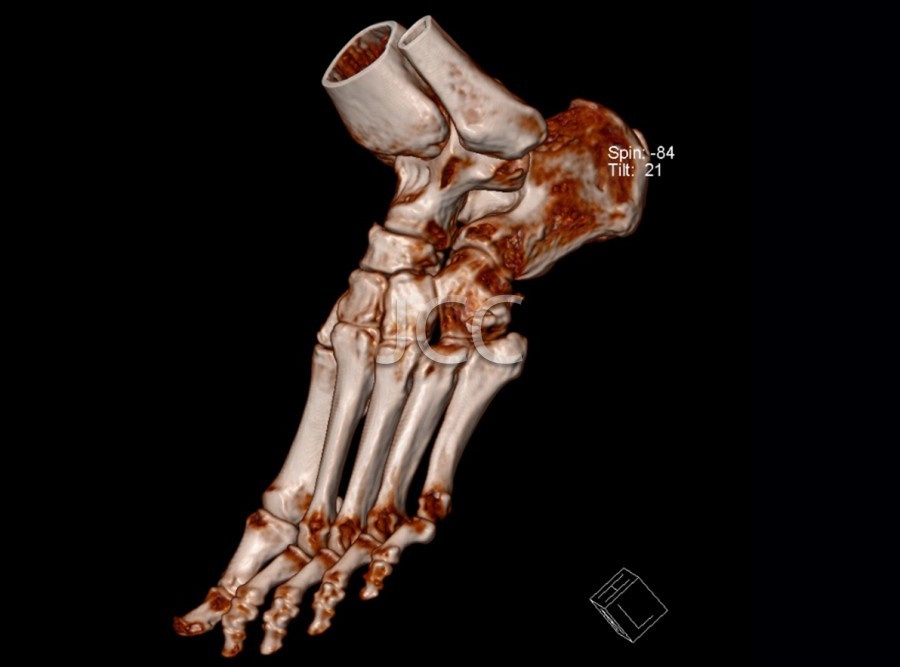
Exams - JCC - João Carlos Costa Diagnóstico por Imagem from www.jcc.pt Ct scans help doctors diagnose and treat medical conditions such as pancreatic cancer. What is a cat scan? A cat (computed axial tomography) scan animation is included because seismic tomography is often compared to cat scans. It is a painless test that takes pictures of the inside of the body. The personnel that perform ct scans are called radiographers or radiology technologists. A computed tomography (ct or cat) scan allows doctors to see inside your body. This can provide structural information about the brain, although it is of a lower resolution than magnetic resonance imaging (mri). Ct scans are especially good for showing bone, soft tissue, and blood vessels.
Or axial, images (often called slices) of the brain.
It enables the doctors to watch vital organs, identify blockages and growths and diagnose signs of diseases without doing surgery. It is sometimes called computerized tomography or computerized axial tomography (cat). If you are having computed. Computed tomography (ct) imaging is also known as computed axial tomography (cat), imaging. A ct scan or computed tomography scan (formerly known as computed axial tomography or cat scan) is a medical imaging technique used in radiology to get detailed images of the body noninvasively for diagnostic purposes. Or axial, images (often called slices) of the brain. Before a ct scan, doctors give patients a contrast dye as a drink or iv. Computerized axial tomography (cat) adalah sebuah penggambaran medis yang menggunakan tomografi melalui proses geometri yang menghasilkan gambar tiga dimensi dari bagian dalam sebuah objek dari satu seri besar gambar sinar x dua dimensi dalam satu putaran axis Computerized axial tomography (cat) is an imaging test used to examine the body's internal structures. This appears to be especially true in children. Ct was conceived by william oldendorf and developed independently by godfrey newbold hounsfield and allan macleod cormack, who shared a 1979 nobel prize for. Ct scans are especially good for showing bone, soft tissue, and blood vessels. It is a painless test that takes pictures of the inside of the body.

_scan_aboard_the_Military_Sealift_Command_(MSC)_hospital_ship_USNS_Mercy_(T-AH_19).jpg)